Flickering light causes responses in the brain at the flicker frequency. This creates the so called Steady State Visually Evoked Potentials (SSVEPs). These responses can be seen in the EEG at frequencies ranging from 1 to 100 Hz (or more), but show higher amplitudes in resonant frequency bands (alpha, beta, gamma etc.) due to an interaction with the preferred frequency of natural oscillations in the brain. SSVEPs have an excellent signal‐to‐noise ratio, and give us the ability to probe specific frequency bands in the cortex.
I am developing a method for producing SSVEPs using adapted Liquid crystal display active 3D glasses (LCD glasses), which are commonly used for 3D TV (see Mobile EEG). Here we adapt this technology to flicker at other frequencies, and with both eyes receiving the same flicker phase, and synchronise with mobile EEG.

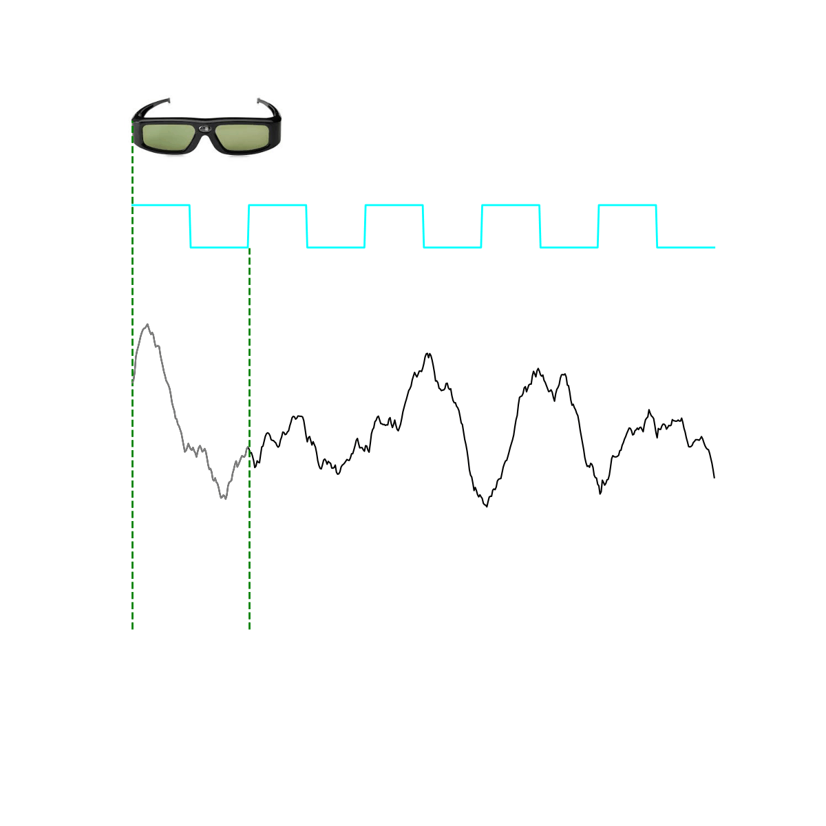
Segments of EEG data can be time-locked to the dark/light of the LCD glass and averaged to make the SSVEP
Using this method SSVEPs can be generated from anything the participant is looking at in the real world; not limited to pictures on a screen. This is important because visual cognitive neuroscience is generally focused on pictures and stimuli presented on screens, the assumption is that the neural correlates of pictures of visual scenes and faces overlap with neural correlates of real places and real things with which we can interact with. Natural stimuli are likely to generate responses that more accurately reflect the functioning of the brain in ordinary life.
A further advantage is that, unlike SSVEPs on a screen which are limited to multiples of the refresh rate, here we can create SSVEPs at any frequency. For example we can do a frequency sweep through the gamma range, or the alpha range and track how the signal resonates with (or entrains) the underlying neural circuits. The amplitude of the SSVEP is largest when the flicker frequency matches the frequency bands of the brain’s natural oscillations. An ongoing debate in the field is whether this is due to entrainment of the brains natural oscillations, or due to resonance with the preferred frequency of the neural circuits.
Here is my resting brain activity (in blue) showing a dominant alpha rhythm at about 11 Hz. In green are SSVEPs: responses to visual flicker at different frequencies from 5 to 15 Hz in steps of 0.5 Hz (i.e. 5, 5.5, 6, 6.5 … etc.).

Pilot data shown below demonstrates that SSVEPs of real world scenes can be generated at a range of gamma frequencies. !!! NOTE: this is just pilot data, n = 1 (i.e. me) !!!
LCD glasses were set to flicker at frequencies from 20 to 56 Hz in steps of 2 Hz, for 1 minute each. Red and blue lines indicate two different visual scenes: a relatively empty scene with a white wall (blue) and a more “busy” visual scene with a bookshelf, desk etc. (red).

Highly similar, and therefore reliable, SSVEPs can be generated throughout the gamma range; the amplitude follows the same broad shape despite the different visual scene. Of interest is the phase/waveform shape of SSVEPs between 20 and 26 Hz which is highly similar in early visual electrodes (O1), but becomes sensitive to differences in the visual scene over parietal electrodes. The SSVEP amplitude peak in the traditional gamma range is 48 Hz at O1 but 54 Hz at P4 (the frequency where O1 shows a sharp drop in amplitude), this indicates resonance with neuronal circuits at different optimal frequencies. Note the different scale; SSVEPs at occipital electrodes are larger.